Each species of Eimeria involved in avian coccidiosis has certain specific characteristics, including where they are located in the intestine and the lesions that each of them causes. Such lesions allow us to diagnose and identify the species involved and, together with other coccidiosis symptoms, confirming the suspect of a disease outbreak.
In this last article of the series (link to part 1 and part 2), we are going to review the intestinal lesions produced by two species of Eimeria, E. necatrix and E. brunetti, as well as severity scorings based on the scale proposed by Johnson&Reid in 1970. As mentioned, scoring lesions is essential to make a right diagnosis of coccidiosis disease.
Lesion score of Eimeria necatrix
Eimeria necatrix affects long-living birds. The lesions are located in the lower and upper mid-intestine, and also in the rectum and caeca in severe cases. Four are the possible lesion scores of affectation, which we will detail below:
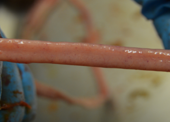
Lesion score 1: small scattered petechiae and white spots are easily seen from the serosal side; little if any damage apparent on the mucosal surface.
Lesion score 2: numerous petechiae on the serosal surface can be observed; a slight ballooning confined to the mid-gut area may be present.
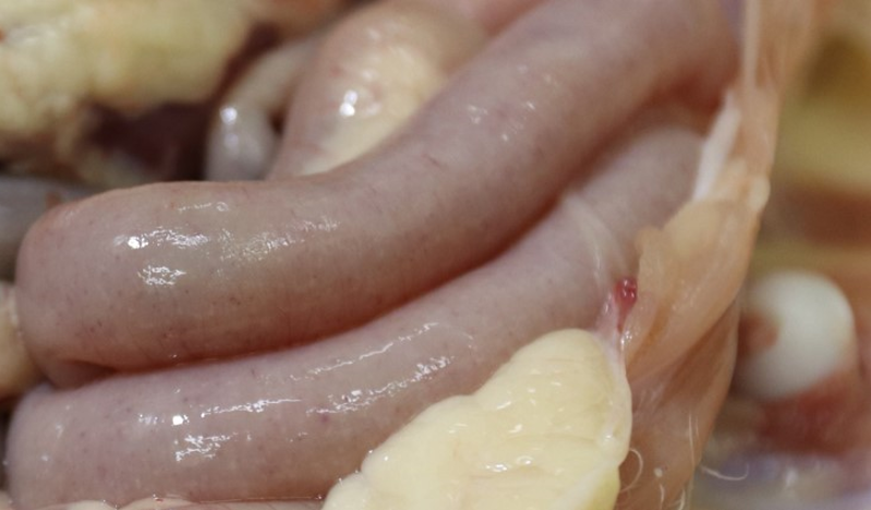
Lesion score 3: an extensive hemorrhage into the lumen of the intestine happens; the serosal surface is covered with red petechiae and/or white plaques (popularly known as “Salt and pepper”). The serosal surface is rough and thickened with many pinpoint hemorrhages. Normal intestinal contents are lacking; ballooning extends over the lower half of the medium intestine.
Lesion Score 3 by Eimeria necatrix, typical “salt and pepper”.
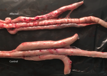
Lesion score 4: extensive hemorrhage is giving the intestine a dark colour; the intestinal contents consist of red or brown mucus. Ballooning may extend throughout much of the length of the intestine. Characteristic putrid odour. Dead birds are scored as 4.
Lesion score of Eimeria brunetti
Eimeria brunetti affects long-living birds. The lesions are located in the lower mid-intestine, rectum and caeca. Four are the possible lesion scores, which we will detail next:
Lesion score 1: no gross lesions are observed. In the absence of distinct lesions, presence of parasites may go undetected unless scrapings from suspicious areas are examined microscopically.
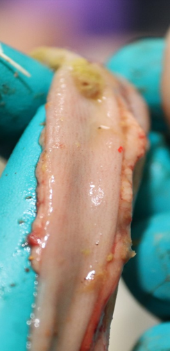
Lesion score 2: the intestinal wall may appear grey in colour. The lower portion may be thickened and flecks of salmon-coloured material sloughed from the intestine are present.
Lesion Score 2 by Eimeria brunetti.
Lesion score 3: the intestinal wall is thickened and a blood-tinged catarrhal exudate is present. Transverse red streaks may be present in the lower rectum and lesions occur in the caecal tonsils. Soft mucus plugs may be present in this latter area.
Lesion score 4: an extensive coagulation necrosis of the mucosal surface of the lower intestine may be present. In some birds, a dry necrotic membrane may line the intestine and caseous cores may plug the caeca. Lesions may extend into the middle or upper intestine. Dead birds are scored 4.
BIBLIOGRAPHY:
- Johnson J., Reid W.M., 1970. Anticoccidial drugs: lesion scoring techniques in battery and floor-pen experiments with chickens. Exp. Parasitology 28(1): 30-6.


