Each species of Eimeria involved in avian coccidiosis has certain specific characteristics, including where they are located in the intestine and the lesions that each of them causes. Such lesions allow us to diagnose and identify the species involved and, together with other coccidiosis symptoms, confirming the suspect of a disease outbreak.
In this article, we are going to review the intestinal lesions produced by two species of Eimeria, E. mitis and E. praecox, not so known as others because they produced mainly subclinical coccidiosis, and they were not described in the scale proposed by Johnson&Reid in 1970.
However, these species can produced gross lesions during severe infestations; HIPRA and other authors (Williams et al, 2009) have developed its own lesion scores for these species, in order to support a right diagnosis of coccidiosis disease.
Lesion score of Eimeria mitis
Eimeria mitis has its relevance in short-living birds. As explained above, it is a species with subclinical effects, so diagnosing it in the field by lesion scoring would not be the preferred technique. It is located principally in the lower mid-intestine and rectum. There are two possible lesion scores, which we will detail below:
Lesion score 1: the intestinal wall presents a normal aspect. It is not thickened. The content is slightly liquid in the medium tract of the intestine.
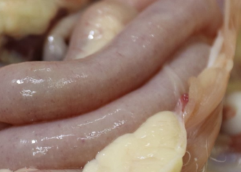
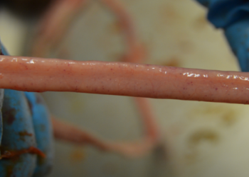
Lesion score 2: the intestinal wall in the tract of the medium lower tract (down the yolk duct) can be slightly thickened and with saw-tooth profile. The content of the lower middle tract has a more creamy aspect. It is observed non-digested feed in the lower part of the gut and in the caeca.
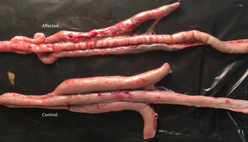
Lesion Score 2 by Eimeria mitis.
Lesion score of Eimeria praecox
Eimeria praecox is relevant in short-living birds. It is also a species with subclinical effects, so diagnosing it in the field by lesion scoring would not be the preferred technique. It is located principally in the duodenum and the upper mid-intestine. Two are the possible lesion scores, which we will detail below:
Lesion score 1: the intestinal wall presents a normal aspect. It is not thickened. The content can be quite liquid in the duodenum and even slightly liquid in the medium tract of the intestine.
Lesion score 2: the intestinal wall in the tract of the duodenum can be slightly thickened. The content is quite liquid. It is observed an increase of whitish mucus in the duodenum and non-digested feed in the lower part of the gut and in the caeca.
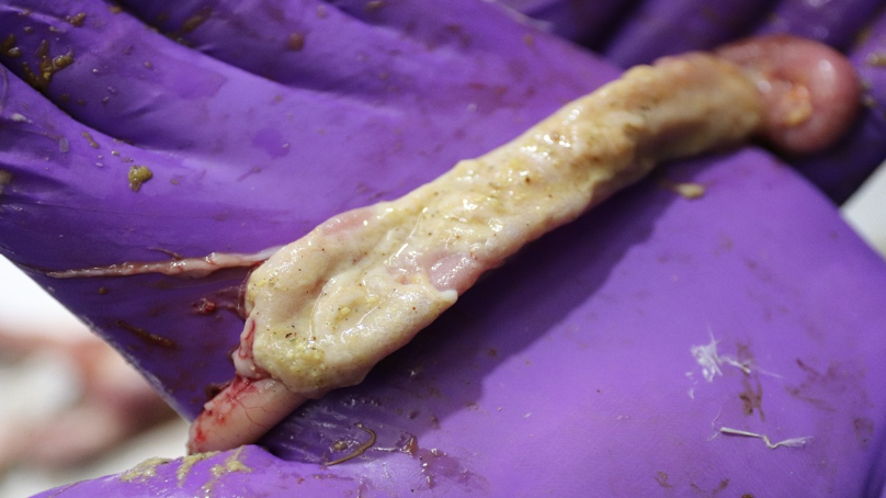
Lesion Score 2 by Eimeria praecox.
BIBLIOGRAPHY:
- Johnson J., Reid W.M., 1970. Anticoccidial drugs: lesion scoring techniques in battery and floor-pen experiments with chickens. Exp. Parasitology 28(1): 30-6.
- Williams R.B., Marshall R.N., Pagès M., Dardi M. and del Cacho E., 2009. Pathogenesis of Eimeria praecox in chickens: virulence of field strains compared with laboratory strains of E. praecox and Eimeria acervulina. Avian Pathology, 38: 5, 359 — 366