Each species of Eimeria involved in avian coccidiosis has certain specific characteristics, including where they are located in the intestine and the lesions that each of them causes. Such lesions allow us to diagnose and identify the species involved and, together with other coccidiosis symptoms, confirming the suspect of a disease outbreak.
In this article and following articles, we are going to review the intestinal lesions produced by different Eimeria spp., as well as severity scorings based on the scale proposed by Johnson&Reid in 1970. As mentioned, scoring lesions is essential to make a right diagnosis of coccidiosis disease.
Lesion score of Eimeria acervulina
Eimeria acervulina affects both short and long-living birds. The lesions are mainly located in the duodenum, but also in the upper and lower mid-intestine. Four are the possible lesion scores of affectation, which we will detail below:
Lesion score 1: scattered, white plaque-like lesions containing developing oocysts are confined to the duodenum. These lesions are elongated with the longer axis transversely oriented on the intestinal walls like the rungs of a ladder. They may be seen from either the serosal or the mucosal intestinal surfaces. They may range up to a maximum of 5 lesions per cm2.
Lesion score 2: lesions are much closer together, but not coalescent; Iesions may extend as far posterior as 20 cm below the duodenum. The intestinal walls show no thickening. Digestive tract contents are normal.
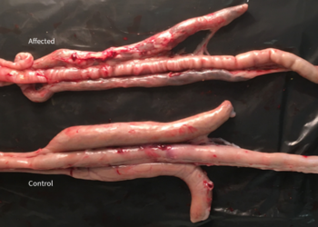
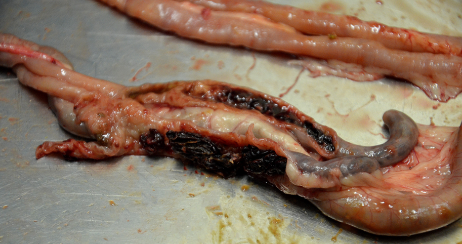
Lesion score 3: lesions are numerous enough to cause coalescence with reduction in lesion size and give the intestine a coated appearance. The intestinal wall is thickened and the contents are watery. Lesions may extend as far posterior as the yolk sac diverticulum.
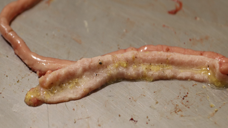
Lesion Score 3 by Eimeria acervulina.
Lesion score 4: the mucosal wall is greyish with colonies completely coalescent. Congestion may be confined to small petechiae or, in extremely heavy infections, the entire mucosa may be bright red in color. Individual lesions may be indistinguishable in the upper intestine. Typical ladder-like lesions appear in the middle part of the intestine. The intestinal wall is very much thickened, and the intestine is filled with a creamy exudate, which may bear large numbers of oocysts. Birds dying of coccidiosis are scored as 4.
Lesion score of Eimeria maxima
Eimeria maxima affects both short and long-living birds. The lesions are located mainly in the mid-intestine, both upper and lower, but can reach the duodenum and rectum in severe cases. Four are the possible lesion scores, which we will detail below:
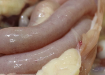
Lesion score 1: small red petechiae may appear on the serosal side of the mid-intestine. There is no ballooning or thickening of the intestine, though small amounts of orange mucus may be present.
Lesion Score 1 by Eimeria maxima.
Lesion score 2: serosal surface may be speckled with numerous red petechiae; the intestine may be filled with orange mucus; little or no ballooning of the intestine may be present; and a thickening of the wall may be observed.
Lesion score 3: intestinal wall is ballooned and thickened. The mucosal surface is roughened; intestinal contents are filled with pinpoint blood clots and mucus.
Lesion score 4: the intestinal wall may be ballooned for most of its length; it contains numerous blood clots and digested red blood cells are giving a characteristic color and putrid odor; the wall is greatly thickened. Dead animals are scored as 4.
Special caution should be exercised in the differential diagnosis with necrotic enteritis, as the lesions caused by that condition are often confused with those caused by Eimeria maxima. Necrotic enteritis causes extensive necrosis and thinning of the intestinal wall. With Eimeria maxima, there are petechiae and thickening of the wall.
Lesion score of Eimeria tenella
Eimeria tenella affects both short and long-living birds. The lesions are located mainly in the caeca, but also in the rectum in severe cases. Four are the lesion scores, which we will detail next:
Lesion score 1: very few scattered petechiae on the caecal wall are observed; there is no thickening of the caecal walls; and normal caecal contents are present.
Lesion score 2: lesions are more numerous with noticeable blood in the caecal contents; cecal wall is somewhat thickened; normal caecal contents present.
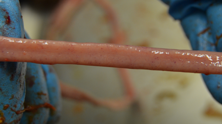
Lesion score 3: large amounts of blood or caecal cores are present; caecal walls greatly thickened; little, if any, faecal contents in the caeca.
Lesion Score 3 by Eimeria tenella.
Lesion score 4: caecal wall is greatly distended with blood or large caseous cores; fecal debris are lacking or included in cores. Dead birds are scored as 4.
BIBLIOGRAPHY:
- Johnson J., Reid W.M., 1970. Anticoccidial drugs: lesion scoring techniques in battery and floor-pen experiments with chickens. Exp. Parasitology 28(1): 30-6.